A new method of image analysis with PEEM and LEEM
Researchers from the Kepler University in Linz along with dr hab. Grażyna Antczak developed a method to analyse images from photoelectron emission microscopy (PEEM) and low energy electron microscopy allowing to determine the type of molecular growth. The results were published in the renowned journal “Ultramicroscopy” in an article titled Standard deviation of microscopy images used as indicator for growth stages (Ultramicroscopy, Vol. 233, March 2022, 113427).
Using this method it is possible to distinguish condensation of solid phases, such as 2D islands or 3D crystallites, from two-dimensional gas phase. Such two-dimensional gas phase consists of molecules that are mobile and diffuse across the whole surface. The individual molecules are too small to be directly observable in PEEM/LEEM images.
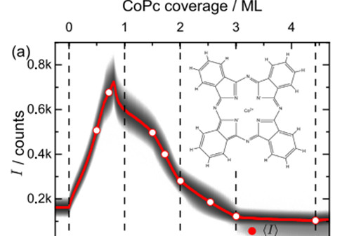
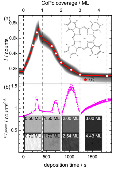
The work discusses how objects below and above the PEEM/LEEM resolution limit affect the average electron yield and its (normalised) standard deviation. The conclusions are illustrated with two experimental examples: molecular growth of cobalt phthalocyanine (CoPc) on Ag (in the figure) and growth of perfluoro-pentacene on the surface of Ag(110). The results demonstrate how spatial and temporal analysis of a series of images can proceed to obtain information about molecular phases that cannot be directly observed on microscopic images.
Added by: Joanna Molenda-Żakowicz
Dean’s representative for promotion and media relations

